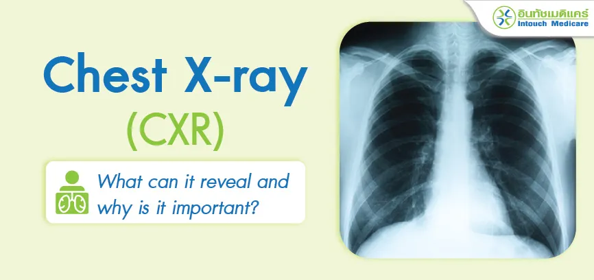
What can a chest X-ray reveal, and why is it important?

A chest X-ray is not just a procedure for detecting abnormalities in the lungs; it also examines the organs in the thoracic cavity, including the lungs, heart, and nearby organs. All of these internal organs play a crucial role in the body’s functioning.
Additionally, interpreting chest X-ray results allows us to see the condition of both normal and abnormal lungs, as well as nearby organs that are not visible to the naked eye, such as white spots in the lungs, Infiltration/opacity, or cardiac shadows. The procedure is also very safe and quick, so patients need not worry.
Interesting Topics

Importance of Chest X-rays
Chest X-rays (CXR) serve as an important screening tool, allowing for the examination of the thoracic area to check for conditions that may not yet exhibit visible symptoms. For instance, it can detect:
Early-stage lung nodules
Enlarged heart size
Ongoing infections (often accompanied by symptoms like fever, cough, and shortness of breath lasting about a week)
In some cases, patients with chronic cough that has been recurring for months, along with weight loss, may be diagnosed with pulmonary tuberculosis through a chest X-ray.

Health Screening Before Employment: Chest X-rays can be used to screen health before starting a job.
In the case of chest X-rays for health screening before employment, they help screen for lung nodules, enlarged heart size, signs of past infections (such as tuberculosis or COVID-19 that may have left pulmonary fibrosis), and any damage that may begin to show on X-ray images before symptoms appear, particularly in patients with a history of chronic smoking.

In cases where the body is already showing symptoms
Chest X-rays are beneficial in identifying the underlying cause, especially in individuals with a persistent cough. They help determine the severity of the condition and provide insight into the potential causes of the symptoms.

In cases of chest trauma from an accident
A chest X-ray can be used to check for rib fractures or conditions like a punctured lung with air leakage (traumatic pneumothorax), among other injuries.
Who Should Undergo Chest X-ray Testing?
Chest X-ray is suitable for screening in various cases, including:
Individuals undergoing pre-employment health screening

Annual health check-ups

People at risk
 , such as those experiencing symptoms like a persistent cough, especially fever, cough with phlegm lasting 3-4 days, chronic cough, heavy smokers (including frequent exposure to secondhand smoke), heart disease patients, and those working in polluted or toxic environments, such as mines or areas with hazardous substances.

Change into the gown provided by the clinic and remove any jewelry, such as necklaces, earrings, belts, or metal accessories. (For women, this includes removing the bra and tying hair up away from the neck area.)
Stand in front of the X-ray machine, where a technician will assist with positioning before the X-ray is taken.
Wait for the technician to give a signal. The technician will count "1, 2, 3" and instruct the patient to take a deep breath and hold it for 3-5 seconds while the X-ray is being taken. The technician will then inform the patient when they can breathe normally again.
After the X-ray procedure is complete, the patient can change back into their clothes and wait to receive the results from the doctor. The images and interpretations will be ready in just a few minutes after the examination.

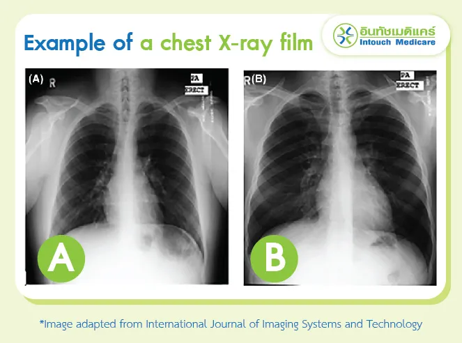
Normal lungs
When interpreting an X-ray film of normal lungs, the following observations can be made: there are no spots, no fibrosis, no fluid in the lungs, and no pneumothorax.
In the image, spot (A) indicates normal lungs, while spot (B) could represent either normal lungs or cardiomegaly. It is essential to consider the patient's body weight as well.

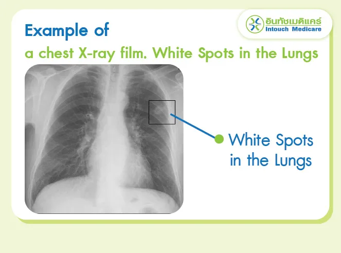
White Spots in the Lungs
In cases where the X-ray results show white spots in the lungs, particularly in abnormal positions, it may indicate an early-stage tumor or signs of a previous tuberculosis infection. The presence of white spots in the lungs depends on the patient's history and symptoms, and a physical examination is also required.

Abnormal Lungs
Abnormal lung conditions can be found in several examples, such as:
infiltration or opacity in the lungs: If accompanied by symptoms like fever, cough, or sputum, this may result from an infection in the pleura or pneumonia, among others.
Masses in the lungs or increased fibrosis: This may be associated with long-term smoking or exposure to secondhand smoke.
Note: All of these are just examples. Every detail must be correlated with the patient's history and physical examination, as it varies for each individual.
Frequently Asked Questions
Should I refrain from eating?
Answer: For a chest X-ray, there is no need to avoid eating. Patients can eat normally.
Why should I avoid wearing jewelry or clothing with metal during the procedure?
Answer: Metal can interfere with the details of the X-ray film. When the radiation is taken, the white shadow of the metal can obscure the lung tissue or the silhouette of the heart (cardiac silhouette).
Are there any side effects?
Answer: The chest X-ray has very few side effects because the amount of radiation used is low, making the risk of developing cancer extremely low, less than 1 in a million.
How long does it take to wait for results?
Answer: The wait time is not long, usually just a few minutes after the X-ray, depending on the symptoms and history of the person being examined.
Does chest X-ray affect pregnancy? How?
Answer: The effect is minimal. Research shows that radiation exposure poses a risk to the fetus only after accumulating more than 70,000 X-ray procedures. Moreover, if the X-ray is performed after the 17th week of pregnancy, the risk is almost negligible.
Why is a chest X-ray necessary before surgery?
Answer: One important reason for having a chest X-ray before surgery is to screen for tuberculosis. In many cases, if a breathing tube is inserted during surgery, it can spread tuberculosis and pose risks to the surgical and anesthesia teams, as well as screen for lung infections and heart size prior to the operation.
Conclusion
A chest X-ray is a diagnostic procedure for examining the thoracic organs. It is a valuable medical test that is quick, provides rapid results, is safe, and offers important details for effective treatment planning.

Moreover, the current risk of PM2.5 pollution is increasing significantly. While there is no definitive research indicating that it raises lung cancer risk, emerging data suggest a "possible" increase in lung cancer risk over time.
![]()
"Therefore, even if there are no symptoms, having the opportunity to undergo a chest X-ray screening can be beneficial for an initial health check, including early detection of nodules in the lungs. If detected early, treatment can begin promptly, preventing further progression."![]()
- Panmai Premashthira, MD. General Practitioner at the clinic. -
references
X-Rays, Pregnancy and You, FDA
Chest X-Ray, Johns Hopkins University School of Medicine
Alveolar deposition of inhaled fine particulate matter increased risk of severity of pulmonary tuberculosis in the upper and middle lobes
For more info and to make an appointment.
Hot Line 081-562-7722
Composer: Panmai Premashthira, MD.
Last edited : 27/09/2024
Images may be used without prior permission exclusively for educational or informational purposes, as long as proper credit is given to intouchmedicare.com
